Coronavirus testing kits that use RNA imaging on the way from Canada


A new hope in the battle against coronavirus is on the way from a team of researchers at Canada’s Simon Fraser University, in Vancouver.
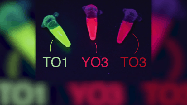
It’s called “Mango”, so-named for its bright, fluorescent color and it uses RNA imaging to detect pathogens such as the coronavirus.
Mango can detect individual RNA molecules within a living cell. The system uses an RNA aptamer (a peptide molecule that binds to a specific target molecule) that attaches to a florescent dye. The dye illuminates the RNA molecules, which makes it much easier for the researchers to see.
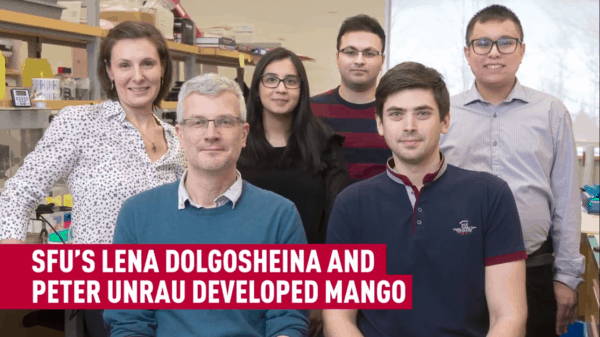
The research was led by Peter Unrau, a professor of molecular biology and biochemistry and was published in the journal Nature Communications on March 9.

“We are made of molecules so when something goes wrong within a cell it happens at the molecular level, Unrau explained. “We are using the Mango system as a catalyst, to allow us to not only extend fundamental research questions but also to detect pathogens like the coronavirus, faster and more efficiently.”
Of course Mango can be used for a variety of diseases.
“Mango technology is state of the art and the development of effective cures for cancer and other diseases demand better imaging methodologies to rapidly learn how cells work in detail,” Unrau added.
Coronavirus sufferers can only hope a solution can come about as quickly as this initiative did.
Funding for the project at SFU came from the Canadian Institutes of Health Research, which on February 20 launched a rapid research funding opportunity that partnered with Genome Canada to provide $6.75-million to contribute to the global response to the 2019-nCoV outbreak, to enhance collaboration of efforts to stop the spread of the virus and to inform a public health response at the national and international levels.
What is RNA Imaging?
Writing for Labome in 2016, researcher Pankaj Kumar provided an overview of various RNA imaging techniques.
“The ability to visualize specific RNA in situ is essential to understand the regulatory mechanisms of gene expression, RNA processing, and splicing,” Kumar wrote. “An improved understanding of these approaches is also necessary in order to identify new drug targets, as well as to create particular gene expression patterns. A variety of protocols have been developed to measure the expression levels of various RNA species, using either fixed-cell or live-cell imaging methods. Despite the fact that much of the knowledge about the spatial distribution of RNA originated from fixed-cell imaging, live-cell imaging strategies provided better possibilities along with added capabilities for real-time monitoring of RNA transport into living cells. These live-cell imaging studies have dramatically improved our understanding of the role of RNA dynamics in various cellular functions. Nevertheless, a range of newer experimental approaches and/or protocols in RNA imaging technologies are continuously evolving.”
Below: Researchers to develop coronavirus tests using SFU-invented RNA imaging technique.
Nick Waddell
Founder of Cantech Letter
Cantech Letter founder and editor Nick Waddell has lived in five Canadian provinces and is proud of his country's often overlooked contributions to the world of science and technology. Waddell takes a regular shift on the Canadian media circuit, making appearances on CTV, CBC and BNN, and contributing to publications such as Canadian Business and Business Insider.
