Study finds software glitch in 40,000 fMRI results

The role of functional magnetic resonance imaging (fMRI) in medical research has been called into question as researchers uncover a significant flaw in the statistical methods and software commonly used to produce data on brain activity and may have affected fMRI results.
In a new paper published in the journal PNAS, Anders Eklund of Linköping University in Sweden concluded that fMRI studies consistently produce false positive results -upwards of 70 per cent of the time- a problem that potentially puts much of the research in neurology conducted over the past twenty years on shaky ground.
“In theory, we should find 5% false positives (for a significance threshold of 5%), but instead we found that the most common software packages for fMRI analysis (SPM, FSL, AFNI) can result in false-positive rates of up to 70%,” say the study’s authors, who along with Eklund include Hans Knutsson also of Linköping University and Thomas E. Nichols of the University of Warwick in the U.K. “These results question the validity of some 40,000 fMRI studies and may have a large impact on the interpretation of neuroimaging results.”
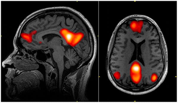
fMRI works by mapping the blood flow in the brain -and hence an image of neural activity- by sending pulses of radio waves through a body (in this case, the brain) surrounded by a strong magnetic field. Radio waves interact with the subatomic nuclei within the brain and cause the nuclei to send out corresponding signals which are then captured by the fMRI’s imaging software. Since its inception over 25 years ago, fMRI has been central to a wide spectrum of research in neurology, valued for its ability to precisely measure blood flow to various regions of the brain.
Eklund and his colleagues analyzed fMRI results from 499 healthy people’s brains in order to find out how often fMRI scans produce false positive results -meaning the brain imaging indicates neural activity trends where the healthy person’s brain is actually at resting state.
Surprisingly, the team found there to be up to a 70 per cent chance that the fMRI results would produce false positives and indicate brain activity where there wasn’t any -far beyond the five per cent error normally expected, say the study’s authors.
“It is not feasible to redo 40,000 fMRI studies, and lamentable archiving and data-sharing practices mean most could not be reanalyzed either,” say the authors. “Considering that it is now possible to evaluate common statistical methods using real fMRI data, the fMRI community should, in our opinion, focus on validation of existing methods.”
Another study published in May of this year in the journal Nature put into doubt the very premise of fMRI-based research, namely, the idea that changes in blood flow in the brain are reflective of localized neural activity.
Researchers looking into neuron activity in response to external stimuli found that blood flow and neural activity are not precisely matched. Instead, so-called “surplus dilation” of blood vessels commonly occurs even without local brain cell activity.
The study’s authors say their work indicates that fMRI’s can at best create “blurred” representations of underlying neural activity rather than a precise detailing of brain functioning.