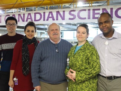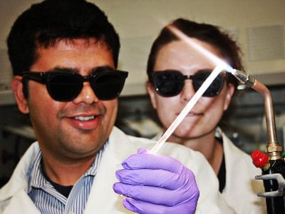

A team of researchers from the University of Saskatchewan’s Canadian Light Source has developed a novel multiple energy X-ray imaging (MEI) system for biomedical imaging, using two or more X-ray energies to visually distinguish and segment contrasting materials based on their spectral absorption, in order to increase diagnostic accuracy.
The team’s research, titled “Multiple energy synchrotron biomedical imaging system”, was published by the Institute of Physics (IOP) in Medicine and Biology.
“We have been working on imaging, including functional imaging, at energies around the K-edge of an element – such as iodine, barium or xenon. Functional imaging is quite powerful for biomedical applications,” said co-author Bassey Bassey to IOP’s medicalphysicsweb site. “Since we developed the technology for preparing such beams, and with the push for clinical imaging systems with dual-energy capability, we explored imaging over a wider energy range. We can easily cover a number of K-edges of elements (four in this paper). We can also use this energy range to discriminate bone from water, which is challenging.”
The elements that Bassey refers to are commonly used as contrast agents in biomedical and clinical imaging.
The Biomedical Imaging and Therapy (BMIT) facility is dedicated to unsolved medical and biological problems, with a concentration on life sciences research at the Canadian Light Source synchrotron, which provides researchers access to two beamlines to facilitate experiments with potential clinical applications in imaging and radiation therapy.
Bassey’s team used the facility’s bend magnet beamline to develop a novel multiple energy X-ray imaging system that has the potential to expand the application of tissue differentiation, and the ability to separate tissue types to improve image contrast and diagnostic accuracy.
The beamline consists of a high vacuum system to transport the X-ray beam, beginning with a source inside the synchrotron’s storage ring, using shutters, slits, filters and monochromators to prepare the beam, and an array of possible instruments, including positioners, stages, detectors to facilitate experiments.
To demonstrate material decomposition, the team used the MEI system to image an object comprising sodium iodide (NaI) solution; xenon gas; caesium chloride (CsCl) solution; a mixture of NaI and barium chloride (BaCl2) solutions; BaCl2 solution; and simulated bone.
Using an algorithm, the researchers successfully decomposed six materials (iodine, xenon, caesium, barium, bone and water) in terms of their projected concentrations.
The Canadian Light Source is a football field sized synchrotron facility housed on the University of Saskatchewan campus in Saskatoon.
The CLS launched in 2004, on completion of construction that began in 1999, and today has over 200 employees and has hosted over 2,500 researchers from various academic and industrial backgrounds, and facilitated countless experiments into cancer research, drug design, solar panel technology, improving the efficiency of computer chips, and a host of other technological applications.
The CLS offers analytical services unavailable anywhere else in Canada, including advanced X-ray imaging, non-destructive testing, in-situ investigations and bulk and surface investigations for the Mining and Environment, Pharmaceuticals, Oil and Gas, Aerospace and Energy Storage sectors.
Leave a Reply
You must be logged in to post a comment.


 Share
Share Tweet
Tweet Share
Share


Comment